
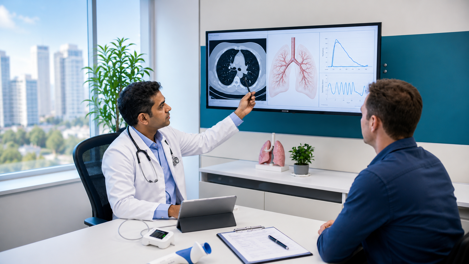
Pulmonology Procedures
Interventional Pulmonology
▲Interventional pulmonology is an emerging subspecialty within pulmonary medicine that focuses on diagnosing and treating conditions affecting the lungs and chest using minimally invasive techniques. These procedures often involve endoscopy and other advanced tools, reducing the need for traditional surgery. Pulmonologists with specialized training in interventional pulmonology perform these procedures, alongside cardiothoracic and other surgeons who routinely incorporate these techniques into their practice. By offering less invasive alternatives, interventional pulmonology helps improve patient outcomes, shorten recovery times, and enhance overall care for respiratory conditions.
Interventional pulmonology includes several advanced procedures to diagnose and treat lung and chest conditions. Flexible bronchoscopy is a common procedure where a thin, flexible tube is inserted through the mouth or nose to examine the airways. Bronchoalveolar lavage involves injecting sterile fluid into the lungs and retrieving it for diagnostic testing. Lung and lymph node biopsies help detect cancer, infections, and other diseases by collecting tissue samples. Airway stents (bronchial stents) are used to open blocked or narrowed airways, while balloon bronchoplasty expands constricted airways using an inflatable balloon. Rigid bronchoscopy, performed under general anaesthesia, allows for complex interventions, including foreign body removal. Pleuroscopy provides a direct view of the lung’s outer surface for diagnosing pleural conditions. Thoracentesis involves draining excess fluid from around the lungs to relieve symptoms and aid diagnosis.
These procedures offer a safer, minimally invasive alternative to traditional surgery, often resulting in shorter recovery times. However, for certain lung conditions, surgery remains the most effective approach for diagnosis and treatment. The choice between interventional procedures and surgery depends on the specific condition, severity, and individual patient needs, emphasising the importance of a tailored treatment plan guided by medical expertise.
Lung Transplant
▼A lung transplant is a surgical procedure that replaces a diseased or failing lung with a healthy one, typically from a deceased donor. It is considered when medications and other treatments are no longer effective. Depending on the condition, a single or double lung transplant may be performed, and in some cases, the lungs are transplanted along with a donor heart. Preparing for a lung transplant begins well in advance, sometimes weeks, months, or even years before receiving a donor's lung.
After surgery, patients spend several days in the ICU, supported by a mechanical ventilator and chest tubes to drain fluids. Following discharge, frequent monitoring over three months is essential to track lung function and prevent complications, requiring regular tests, imaging, and specialist checkups, often near the transplant centre. Long-term adjustments include:
- Immunosuppressant therapy to prevent organ rejection.
- A healthy lifestyle with structured pulmonary rehabilitation to improve breathing and overall function.
- Emotional support, as the physical and emotional demands of a transplant can be overwhelming.
While it is a complex procedure with potential risks, a lung transplant can significantly enhance both health and quality of life.
Bronchoalveolar Lavage (BAL)
▼Bronchoalveolar lavage (BAL) is a widely used and relatively safe diagnostic procedure for assessing lung diseases. It is particularly valuable when a patient’s clinical history, physical examination, routine lab tests, pulmonary function tests, and imaging do not provide a clear diagnosis. Unlike sputum analysis, BAL enables targeted sampling of the lower respiratory tract, minimising contamination from the upper airways and improving diagnostic accuracy. This procedure plays a crucial role in identifying infections, inflammatory conditions, and other lung disorders, aiding in more precise and effective treatment planning.
Flexible Bronchoscopy
▼Bronchoscopy is a vital procedure that allows doctors to examine the lungs and airways, often performed by a pulmonologist. A flexible bronchoscope helps diagnose conditions like persistent cough, infections, or abnormal imaging findings. In cases of heavy bleeding or airway blockages, a rigid bronchoscope may be needed. Beyond diagnosis, bronchoscopy plays a key role in collecting mucus or tissue samples, removing foreign objects, clearing blockages, and even delivering targeted treatments. With its versatility and minimally invasive nature, bronchoscopy is an essential tool in modern respiratory care.
During bronchoscopy, specialised tools can be used to enhance both diagnosis and treatment—from biopsy instruments for tissue sampling to electrocautery probes for controlling bleeding and lasers for reducing airway tumours. Advanced techniques ensure precise biopsy collection for accurate diagnosis. The procedure is typically done in hospital, with the full process, including preparation and recovery, taking about four hours, while the bronchoscopy procedure lasts 30 to 60 minutes.
Pulmonary Endarterectomy
▼Pulmonary endarterectomy (PEA) is the preferred treatment for chronic thromboembolic pulmonary hypertension (CTEPH), aiming to remove obstructive material from the pulmonary arteries. CTEPH is a rare but serious form of pulmonary hypertension (PH) caused by long-term obstruction of the pulmonary arteries due to persistent blood clots. Unlike acute pulmonary embolism, where clots dissolve over time, in CTEPH, they become fibrotic and scar-like, leading to narrowed or blocked arteries, increased pulmonary vascular resistance, and elevated pressure in the lungs. This forces the right side of the heart to work harder, potentially leading to right heart failure if untreated.
Pulmonary endarterectomy is a complex surgery that reduces pulmonary vascular resistance, alleviates pulmonary hypertension (PH), and improves right ventricular function, offering both symptomatic relief and long-term benefits for most patients. However, not all individuals with CTEPH are suitable candidates, as operability assessment is complex and requires expert evaluation. Referral to an experienced CTEPH centre is essential to determine the best treatment approach.
Chest Tube Procedure
▼A chest tube is a plastic tube used to drain air or fluid, such as blood or pus, from the pleural space—the area between the lungs and the chest wall. This buildup can cause lung collapse, requiring intervention. Chest tubes can be inserted during surgery under anaesthesia or at the bedside using a local anaesthetic and sedation. Once placed, chest tubes are connected to a drainage system, which allows fluid to drain passively or with suction assistance.
Patients undergoing the procedure while awake receive local anaesthesia and medication to ease discomfort, though some pain and pressure are common. The placement techniques may vary; some tubes are inserted by spreading chest wall tissues, while others use a guidewire technique. The primary goal of this procedure is to restore normal lung function by draining the pleural space.
Thoracentesis
▼Thoracentesis is a minimally invasive procedure used to remove fluid from the pleural space—the area between the lungs and chest wall. Conditions such as infections, heart failure, or cancer can cause fluid buildup (pleural effusion), leading to breathing difficulties. This procedure helps relieve symptoms and allows for fluid analysis to determine the underlying cause. Typically performed by a surgeon, pulmonologist, or radiologist, thoracentesis takes about 15 minutes.
In some cases, a chest tube or pigtail catheter may be placed for ongoing drainage over a few days. Since it is not a major surgery, recovery is quick, with lower risks compared to more invasive procedures. Patients may experience temporary coughing as their lungs re-expand. Post-procedure imaging, such as an X-ray or ultrasound, ensures proper lung function. The collected fluid is analysed for infection or disease, and results help guide further treatment.
Video-Assisted Thoracic Surgery (VATS)
▼Video-assisted thoracoscopic surgery (VATS) is a minimally invasive technique used to diagnose and treat chest conditions. During the procedure, a thoracoscope (tiny camera) and surgical instruments are inserted through small incisions in the chest wall. The camera transmits live images to a monitor, allowing the surgeon to operate with precision. VATS is used for a variety of procedures, including:
- Biopsy and tissue removal for diagnosing lung cancer, pleural mesothelioma, and other chest cancers.
- Lung surgery, including cancer treatment and lung volume reduction.
- Draining excess fluid or air around the lungs.
- Hyperhidrosis treatment to reduce excessive sweating.
- Esophageal procedures, such as esophagectomy for cancer or hernia repair.
- Thymectomy to remove the thymus gland.
- Surgical interventions involving the heart, ribs, spine, and diaphragm.
As a less invasive alternative to open surgery, VATS typically results in faster recovery, less pain, and reduced complications.
Lobectomy
▼A lobectomy is a surgical procedure to remove a lobe of the lung, most commonly performed to treat lung cancer. It is often curative in early-stage cases, though its effectiveness decreases for larger tumours. This procedure may also be necessary for congenital lung abnormalities, lung damage from infection or radiation, metastatic tumours, or significant bleeding. As a major surgery, lobectomy typically requires a four to five-day hospital stay for open-chest procedures.
However, minimally invasive techniques, such as video-assisted thoracoscopic surgery (VATS) or robotic-assisted thoracic surgery (RATS), offer comparable outcomes with reduced pain, shorter hospital stays (two to three days), and faster recovery. These approaches allow patients to return to daily activities more quickly than with traditional open surgery. Full recovery generally takes at least a month, though minimally invasive methods may shorten this timeframe.
Robotic-Assisted Bronchoscopy
▼Robotic bronchoscopy, or robotic-assisted bronchoscopy, is an advanced, minimally invasive procedure used to biopsy lung nodules for the detection of lung cancer and other lung diseases. While traditional bronchoscopy requires a doctor to manually guide a bronchoscope through the airways, robotic bronchoscopy enhances precision by using a robotic arm controlled from a console.
The robotic system directs a flexible, manoeuvrable catheter equipped with a camera, light, and shape-sensing technology, allowing doctors to navigate tight turns and hard-to-reach areas of the lungs with greater accuracy. This improves access to suspicious nodules that may not be reachable with conventional methods. The procedure is carried out under general anaesthesia, ensuring patient comfort. It is typically an outpatient procedure, with minimal recovery time and a low risk of complications. Robotic bronchoscopy enhances diagnostic accuracy while maintaining safety and efficiency.